
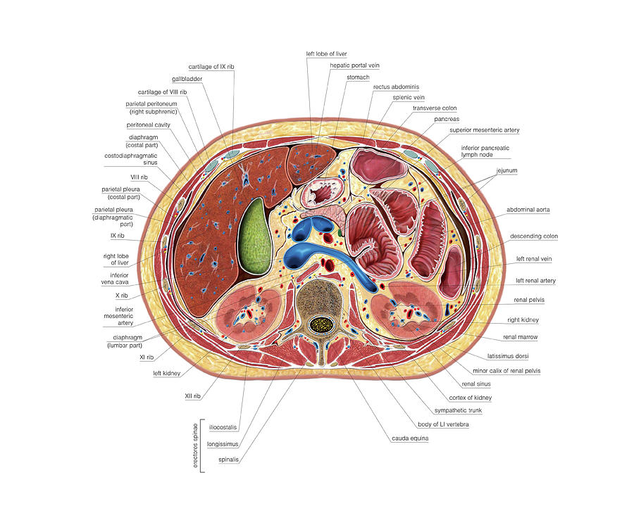
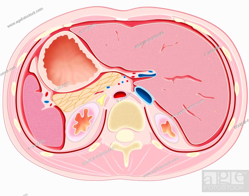
Schematic Cross Section through Abdomen (T12) code 6000.16 Altay Scientific
Cadaveric Preparation. A human donor with appropriate consent for retention and imaging was accepted by the University of Bristol bequest office. Upon arrival the cadaver was thoroughly cleaned, disinfected and embalmed using a formaldehyde based embalming fluid, via injection through the right femoral artery.

Photographic crosssection of the abdomen from the Visible Human Male. Download Scientific Diagram
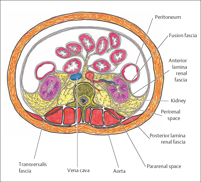
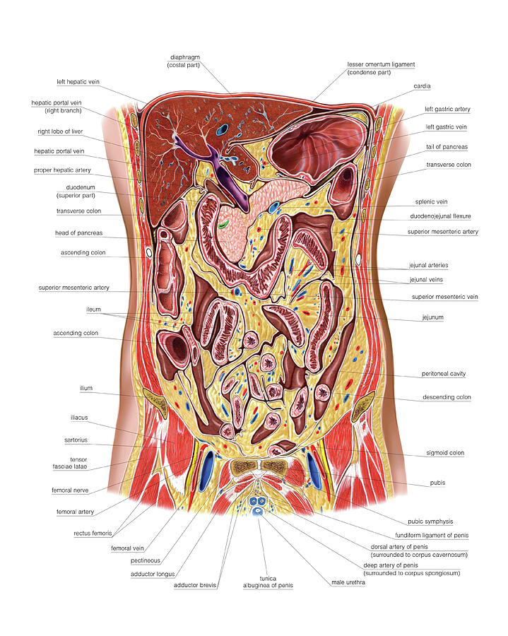
The abdominal wall surrounds the abdominal cavity, providing it with flexible coverage and protecting the internal organs from damage. It is bounded superiorly by the xiphoid process and costal margins, posteriorly by the vertebral column and inferiorly by the pelvic bones and inguinal ligament.. The abdominal wall can be divided into two sections: anterolateral and posterior abdominal walls.

Crosssection illustration of the abdomen at the waist . From left to... News Photo Getty Images
Stomach Cross-section Food enters the body through the mouth. Inside, mechanical breakdown begins immediately as it is chewed and mixed with saliva. This breakdown continues as the food travels.

Abdominal Cross Section (T12) Diagram Quizlet
This session covers Sectional anatomy of Abdomen , Pelvis.Cross Section as well as Mid Sagittal section Sections covered.Gross Specimens compared with CT Sca.
The Abdomen 2 by Asklepios Medical Atlas
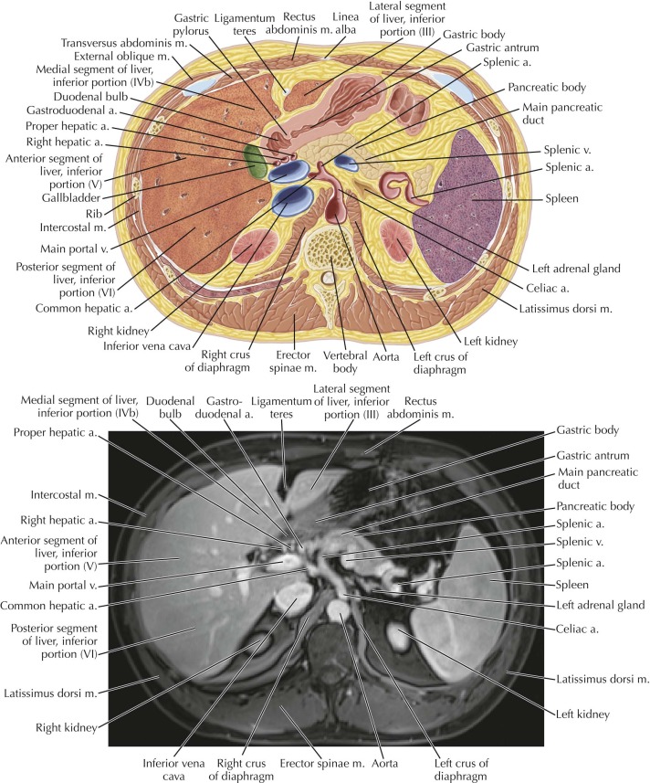
Anatomical structures of the abdomen and pelvis are visible as interactive labeled images. Cross sectional anatomy: MDCT of the abdomen and pelvis An enhanced (portal venous phase - 70 seconds) multidetector computed tomography was performed on a healthy subject in axial plane with coronal and sagittal reformatted images.
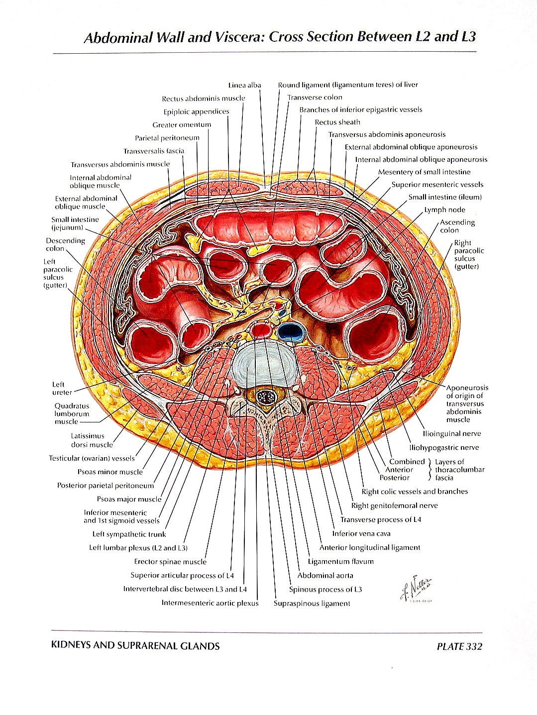
Anatomy Print Abdominal Walls and Viscera Cross Section
The transversus abdominis muscle is the most internal muscle of the anterolateral abdominal wall musculature and its orientation is transverse. It arises from the lateral iliopubic tract, the iliac crest, the lumbodorsal fascia, and the lower six ribs. It fuses with the internal oblique aponeurosis to become the posterior rectus sheath [ 7 ].

cross sectional anatomy abdomen
EXERCISE: Abdomen Cross Section Section 3: Anatomy of the Pelvis (15:19) EXERCISE: Pelvis Cross Section Section 4: Anatomy of Musculoskeletal (29:45) Slides: Anatomy of Chest, Abdomen & Pelvis Week 11: Test (20 Questions) Week 12: CT Artifacts Section 1: Artifacts - Appearance, Cause & The Fix (23:45).
Abdomen Radiology Key
Videos Quizzes Abdomen Peritoneum and peritoneal cavity Stomach Spleen Pancreas Liver and gallbladder Small intestine Large intestine Kidneys, ureters and adrenal glands Pelvis Perineum Urinary bladder and urethra Female reproductive organs Male reproductive organs Blood vessels Innervation Lymphatics Sources Related articles Abdomen and pelvis
Posterior Aspect of the Abdominal Viscera and Retroperitoneum Oncohema Key
Abdominal computed tomography (CT) is a type of medical imaging procedure used to diagnose and monitor internal stomach issues, like cancer, bowel obstruction, and abdominal pain. Radiographers suggest an abdominal CT scan to look for the following: Hernia Cause of a fever Kidney stones Appendicitis Cause of blood in the urine

Axis Scientific Anatomy Model of Abdominal Cross Section at T12 Offers View of Thorax at T12
Crosssection illustration of organs in the abdomen at T12L1, Stock Photo, Picture And Rights
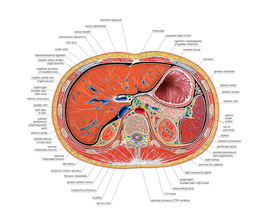
Abdominal cross‐sectional segment of trunk. This cross‐sectional segment is bounded superiorly by a virtual horizontal plane at the level of the junction T8/T9 and inferiorly by a virtual horizontal plane traversing the superior boundary of the iliac crest at the level of the intercristal line (also termed Jacoby's or Tuffler's line), which.
The Abdomen by Asklepios Medical Atlas
Figure \(\PageIndex{2}\): Muscles of the Abdomen - Cross Section. When viewed in cross section the relative positions of the lateral muscles can easily be seen. The tendon of the external oblique passes superficial to the rectus abdominus while the tendon of the transversus abdominus passes deep. The tendon of the internal oblique splits to.
The Abdomen Photograph by Asklepios Medical Atlas Pixels
This video deals with the anatomy of abdominal viscera and walls as they appear in transverse anatomical sections and axial CT sections. The video begins wit.

Transverse Cross Section of Abdomen Diagram Quizlet
Abdomen Cross Section - luc.edu

Schematic Cross Section of Abdomen at Middle T12 Anatomy Liver , Falciform ligament , Superior
anatomical parts. The Visible Human Project® is an outgrowth of the NLM's 1986 Long-Range Plan. It is the creation of complete, anatomically detailed, three-dimensional representations of the normal male human body. Complete cross-sectional images of representative male cadaver. The male was sectioned at one millimeter intervals.

CT scan of abdomen (crosssection view). Download Scientific Diagram
Abdomen - Axial cross section: Omental bursa; Lesser sac, Omental foramen; Epiploic foramen Intestinal tract: 2 illustrations of gross anatomy to introduce the different parts of the digestive tract. Gastrointestinal tract: Oesophagus, Stomach, Small intestine, Large intestine Stomach: anatomical images of the gastric anatomy, from the serous.